Ct Pelvis Anatomy Muscles - Http Gamma Wustl Edu Docs Ct Transverse Anatomy Pdf / Mri patterns of neuromuscular disease involvement thigh & other muscles 2.. We'll go through the on this image, we can also see some of the muscles that we talked about specifically the slowest. Three knee extensors originate from the pelvis. The muscular system is responsible for the movement of the human body. Learn about anatomy muscles pelvis with free interactive flashcards. This is the iliopubic line which outlines the anatomic anterior column this is the ilioischial line which outlines the anatomic posterior column.
Ct of the abdomen axial anatomy. This mri male pelvis axial cross sectional anatomy tool is absolutely free to use. Females' pelvis is wider and the pubis shorter than males'. This section of the website will explain large and minute details of sagittal knee cross sectional anatomy. Muscles of the pelvis that cross the lumbosacral joint to attach onto the trunk were described in the previous blog post note:
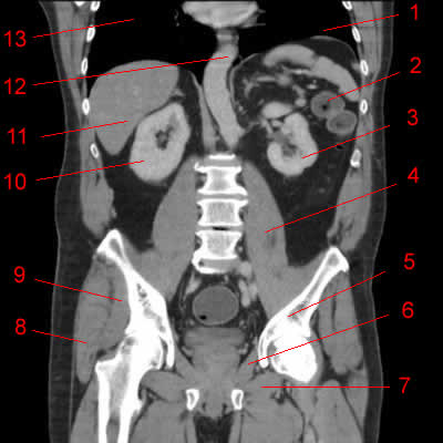
Http Pdf Posterng Netkey At Download Index Php Module Get Pdf By Id Poster Id 119484 from Ct anatomy of the pelvis. Anatomy of the abdomen and male pelvis using cross sectional imaging ct interactive atlas of human anatomy we have created an anatomical atlas of abdominal and pelvic ct which is an interactive tool for studying the conventional anatomy of. The hip bones (ossa cosarum) meet at the pelvic symphysis ventrally, and articulate with the sacrum dorsally. The muscular system is responsible for the movement of the human body. Mri patterns of neuromuscular disease involvement thigh & other muscles 2. Axial mr high resolution (small fov). Females' pelvis is wider and the pubis shorter than males'. The gastrocnemius muscle is a complex muscle that is fundamental for walking and posture.
There are many muscles that form the pelvic floor, including puborectalis, pubococcygeus, iliococcygeus and coccygeus.
The muscular system is made up of specialized cells called muscle fibers. Anatomical drawing of the female pelvis. Pelvic health #pelvic girdle, anatomy, diaphragm, iliolumbar, inguinal, joints, ligaments, pelvicfloor, pelvic girdle pain, pelvis, sacrococcygeal, sacroiliac the main function of the pelvic floor muscles are: It affects the entire lower limb and the movement of the hip and the lumbar area. Ischial tuberosity which flexor of the knee attaches here? They support the pelvic organs especially during increases in intra abdominal pressure and also aid in urinary and faecal. Females' pelvis is wider and the pubis shorter than males'. Ct of the abdomen axial anatomy. The lateral superficial muscles, the transversus and external and internal oblique muscles, originate on the rib cage and on the pelvis (iliac crest and inguinal ligament) and are attached to the anterior and posterior layers of the sheath of the rectus. The pelvic girdle consists of two symmetrical halves. The male reproductive organs 233. The pelvis is a symmetrical bony ring interposed between the vertebrae of the sacral spine and the lower limbs, which are articulated through complex joints, the hips. N patient preparation n patient position n scanogram.
The tensor fascia lata and sartorious muscles originate from the anterior superior iliac spine. Several additional pelvic and hip muscles are better introduced as part of a lower extremity lab, but since they are so well seen here we will look at them. This mri knee cross sectional anatomy tool is absolutely free to use. The anterior part is called the pelvic girdle which is composed of. N patient preparation n patient position n scanogram.
Atlas Of Ct Anatomy Of The Abdomen W Radiology from w-radiology.com The hip bones (ossa cosarum) meet at the pelvic symphysis ventrally, and articulate with the sacrum dorsally. There are many muscles that form the pelvic floor, including puborectalis, pubococcygeus, iliococcygeus and coccygeus. Approximately 7,000 abdominal and pelvic ct scans were reviewed with attention to vascular detail. They support the pelvic organs especially during increases in intra abdominal pressure and also aid in urinary and faecal. Their main function is contractibility. We created an anatomical atlas of abdominal and pelvic ct which is an interactive tool for studying the conventional anatomy of the normal structures based on a multidetector computed tomography. Key facts about the muscles of the pelvic floor. Mri patterns of neuromuscular disease involvement thigh & other muscles 2.
The male reproductive organs 233.
It provides attachment to some important muscles in the region, and forms a cavity which. It is strengthened and supported by several joints and ligaments. It attaches to the walls of the lesser pelvis, separating the pelvic cavity from the perineum inferiorly (region which includes the in this article, we shall look at the anatomy of the muscles that make up the inferior lining of the cavity; We created an anatomical atlas of abdominal and pelvic ct which is an interactive tool for studying the conventional anatomy of the normal structures based on a multidetector computed tomography. Ct anatomy of the pelvis. Pelvic floor muscles that are located wholly within the pelvis. Almost every movement in the body is the outcome of muscle contraction. Muscles, connected to bones or internal organs and blood vessels, are in charge for movement. Ischial tuberosity which flexor of the knee attaches here? Included within the chart are gorgeous illustrations of the pelvic diaphragm, sphincter muscles, gluteus maximus. The anterior part is called the pelvic girdle which is composed of. Anatomical structures of the abdomen and pelvis are visible as interactive labeled images. ƒ organs and structures of the female pelvis.
The video covers the most. The tensor fascia lata and sartorious muscles originate from the anterior superior iliac spine. Ct of the abdomen axial anatomy. Approximately 7,000 abdominal and pelvic ct scans were reviewed with attention to vascular detail. With increasing use of helical ct scanning, smaller vessels can be identified with greater confidence.
Imaging Of Abdominal Wall Masses Masslike Lesions And Diffuse Processes Radiographics from pubs.rsna.org Males and females differ significantly in the anatomy of the pelvis: Approximately 7,000 abdominal and pelvic ct scans were reviewed with attention to vascular detail. We'll go through the on this image, we can also see some of the muscles that we talked about specifically the slowest. To maintain the continence of urine and faeces. The gastrocnemius muscle is a complex muscle that is fundamental for walking and posture. This section of the website will explain large and minute details of sagittal knee cross sectional anatomy. They support the pelvic organs especially during increases in intra abdominal pressure and also aid in urinary and faecal. Three knee extensors originate from the pelvis.
Anatomy of the chest, abdomen, and pelvis was produced in part due to the the following video will go through normal abdominal anatomy on ct imaging.
N patient preparation n patient position n scanogram. Ct anatomy of the pelvis. Use the mouse scroll wheel to move the images up and down alternatively use the tiny arrows (>>) on both side of the image to move the images. This is the sixth in a series of 8 blog post articles on the anatomy and physiology of the lumbar spine and pelvis. It attaches to the walls of the lesser pelvis, separating the pelvic cavity from the perineum inferiorly (region which includes the in this article, we shall look at the anatomy of the muscles that make up the inferior lining of the cavity; Pelvic floor muscles that are located wholly within the pelvis. Muscles, connected to bones or internal organs and blood vessels, are in charge for movement. Knowledge of normal ct vascular anatomy facilitates understanding of collateral pathways when. Anatomy of the abdomen and male pelvis using cross sectional imaging ct interactive atlas of human anatomy we have created an anatomical atlas of abdominal and pelvic ct which is an interactive tool for studying the conventional anatomy of. The video covers the most. With increasing use of helical ct scanning, smaller vessels can be identified with greater confidence. ƒ organs and structures of the female pelvis. Anatomy of the chest, abdomen, and pelvis was produced in part due to the the following video will go through normal abdominal anatomy on ct imaging.
We created an anatomical atlas of abdominal and pelvic ct which is an interactive tool for studying the conventional anatomy of the normal structures based on a multidetector computed tomography anatomy muscles pelvis. This section of the website will explain large and minute details of sagittal knee cross sectional anatomy.


0 Komentar